History
A short history of the light microscope
From hand-ground lenses in seventeenth-century Holland to LED-lit research stands. The handful of ideas that made the modern microscope.
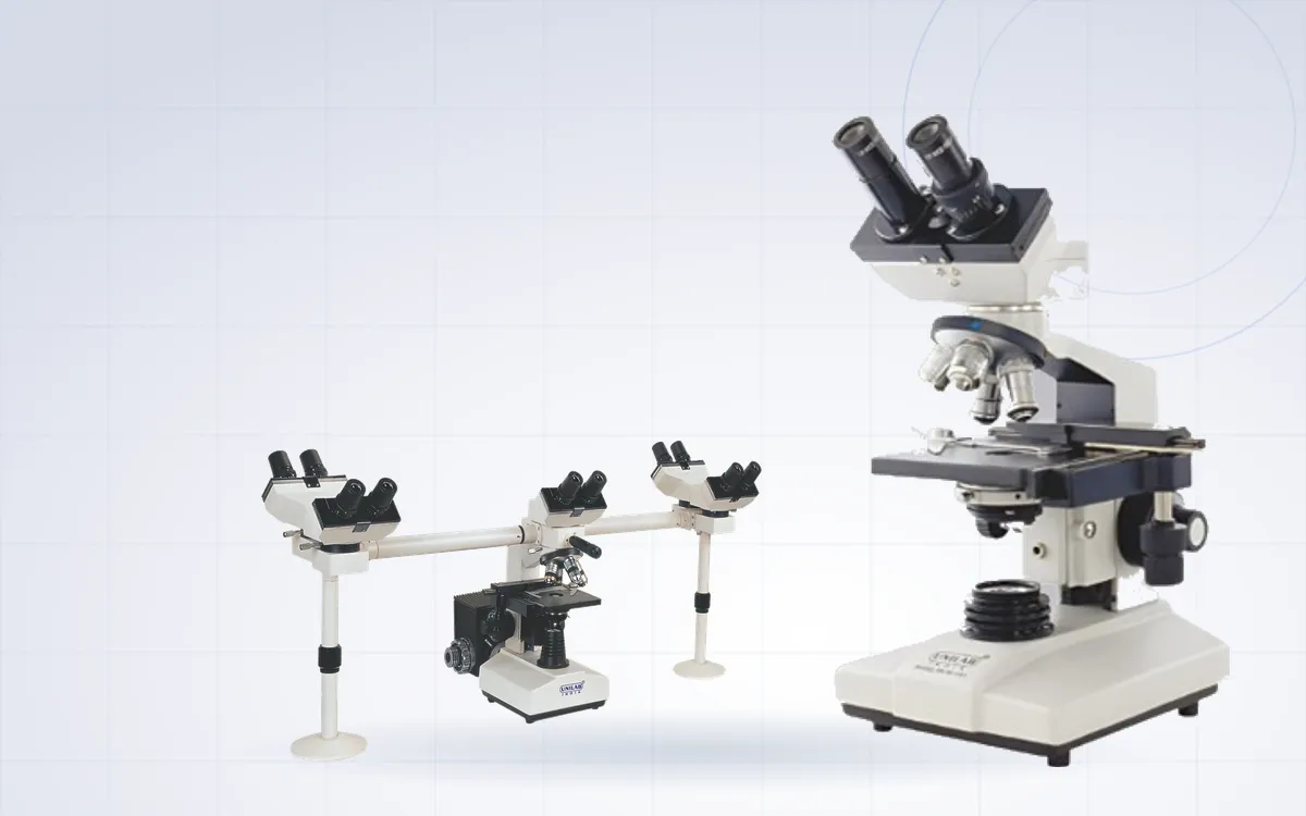
The microscope on a laboratory bench today looks nothing like the brass instruments of the 1600s. Yet almost every part of it can be traced to a few people who solved one specific problem at a time.
The first compound microscopes
Spectacle makers in the Netherlands were combining lenses in tubes by the end of the sixteenth century. Exactly who built the first compound microscope is still debated. What is clear is that the early instruments magnified only modestly and the images were blurred and fringed with colour.
Hooke and Leeuwenhoek
In 1665 Robert Hooke published Micrographia, with detailed drawings of insects, plant tissue and the small compartments in cork that he called cells.
A few years later Antonie van Leeuwenhoek, a cloth merchant in Delft, began making tiny single-lens microscopes of remarkable quality. From the 1670s he described what he saw in letters to the Royal Society in London, including single-celled organisms and, later, bacteria. For a time, his simple microscopes outperformed compound instruments.
Fixing colour
Simple lenses bend different colours by different amounts, so the image has coloured edges. Achromatic lens combinations, pairing two types of glass, reduced this. In the 1830s Joseph Jackson Lister applied careful optical design to microscope objectives, and good achromatic objectives became standard.
Abbe, Zeiss and the theory of resolution
In Jena in the 1870s, the physicist Ernst Abbe working with Carl Zeiss's workshop put microscope design on a scientific footing. Abbe explained how resolution depends on the wavelength of light and the numerical aperture of the objective. He introduced homogeneous oil immersion and, with new optical glass from Otto Schott, apochromatic objectives that corrected colour even more closely.
The numerical aperture printed on every objective today comes from this work.
Even light: Köhler
In 1893 August Köhler described a way of setting up the illumination so the specimen is evenly lit and the image is as sharp as the optics allow. Köhler illumination is still taught to every new microscopist.
Seeing without stains
Many living cells are nearly transparent. In the 1930s Frits Zernike developed phase contrast, which turns small differences in the way light passes through a specimen into differences in brightness. He received the Nobel Prize in Physics in 1953. Fluorescence methods, developed through the twentieth century, added a way to make specific structures glow.
The modern bench microscope
Infinity-corrected optics, plan objectives with a flat field, cool LED illumination, multi-head teaching bridges and integrated digital cameras are the latest steps. The basic layout, an objective, an eyepiece and a carefully controlled light path, is the one that Abbe and Köhler would recognise.
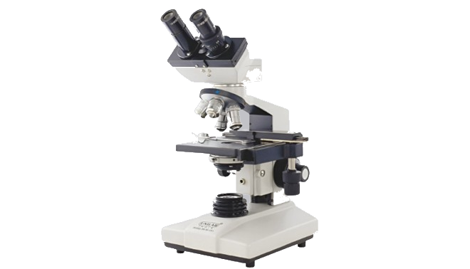
Advance Binocular Research Microscope
Best for: Research and teaching laboratories
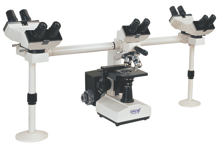
Penta Head Binocular Research Microscope
Best for: Teaching, pathology review and case discussion