Fluorescence microscopy: what a lab needs to get started
Filters, light sources, a dark room and a few working habits. A practical overview for laboratories adding fluorescence to routine microscopy.
Research groups need imaging that can be documented and reproduced, and bench equipment that holds its settings. This shortlist covers fluorescence and cell-culture microscopy as well as sample preparation.
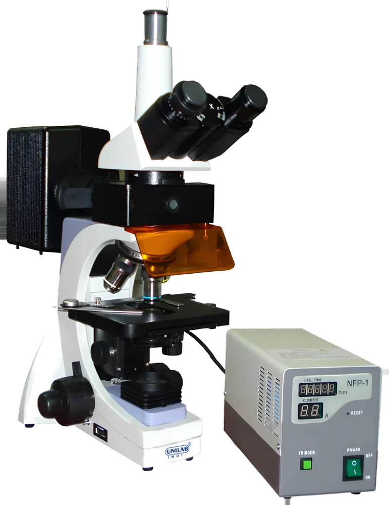
Used to view specimens that emit fluorescence under specific light wavelengths.
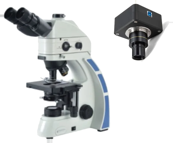
Best for: Fluorescence & imaging
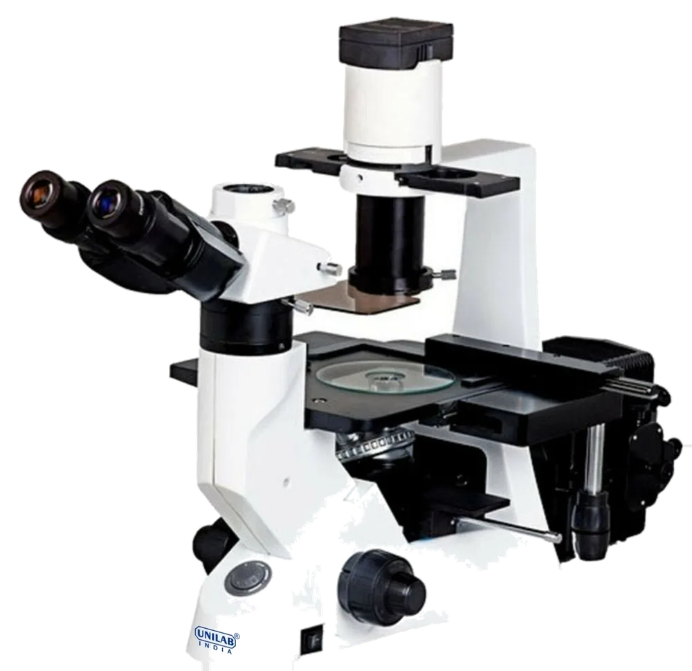
For biological samples(Cell/Tissue Cultures),Morphology(zoology/biology),Veterinary science,Laboratory.
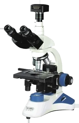
Best for: Trinocular research
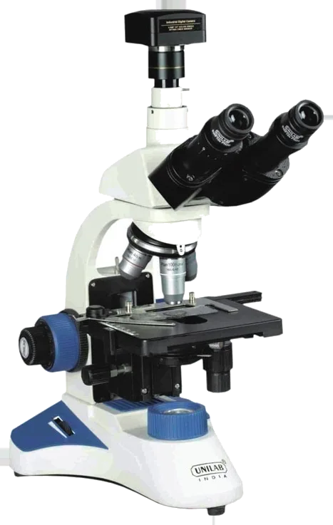
For pathological observation.
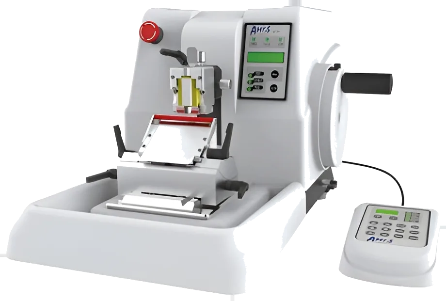
Best for: Rotary microtomes
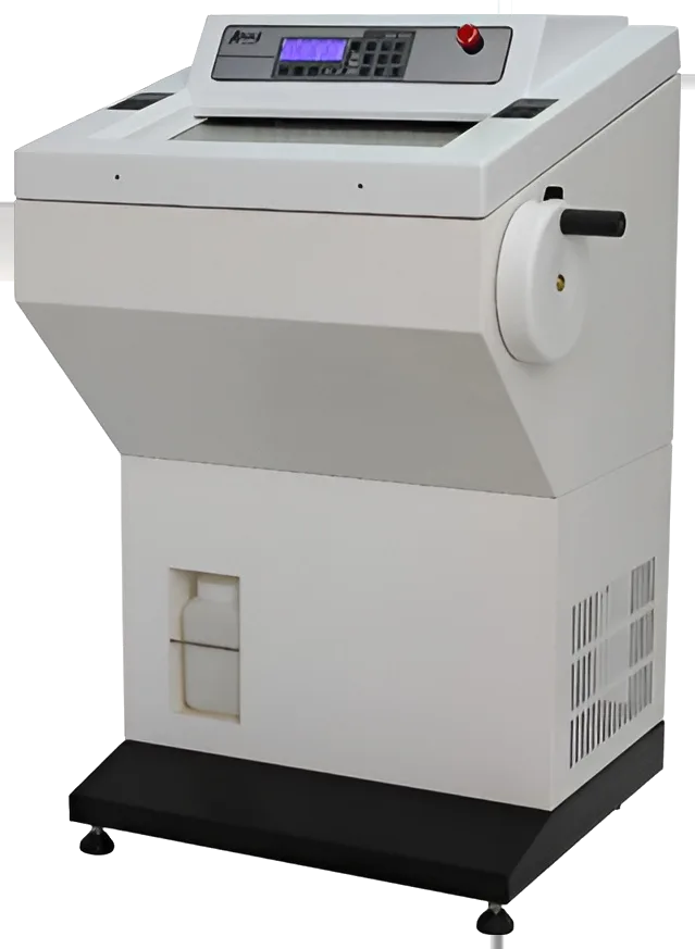
Best for: Cryostats
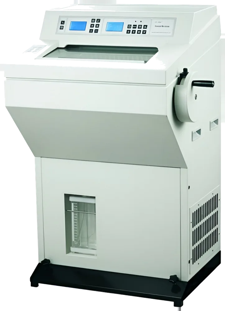
Best for: Cryostats
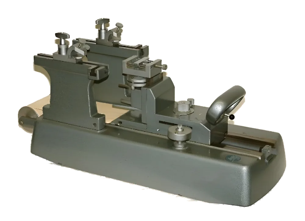
Best for: Specialist microtomes
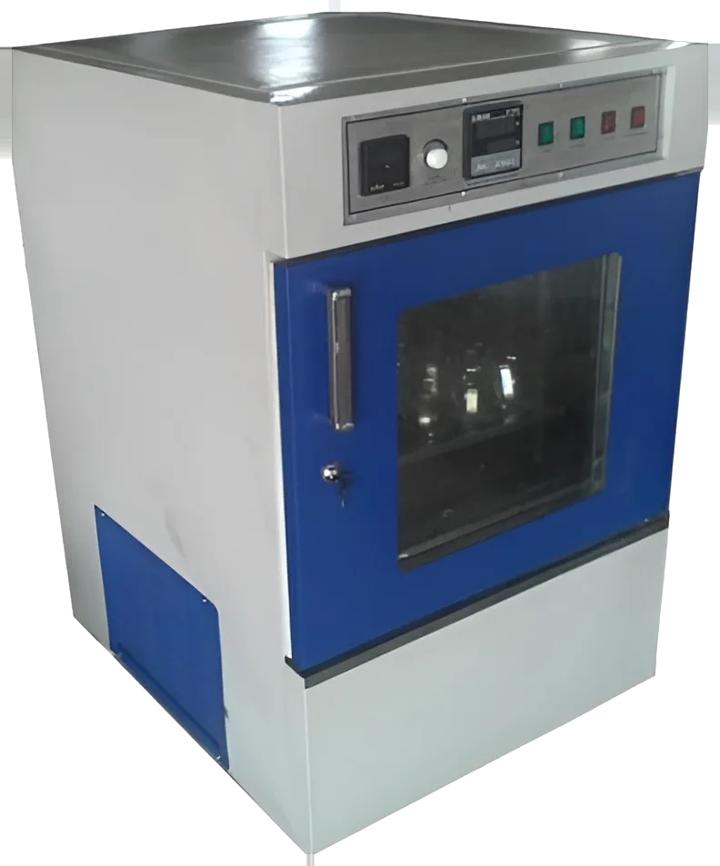
Best for: Incubators
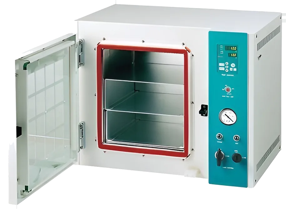
Best for: Ovens
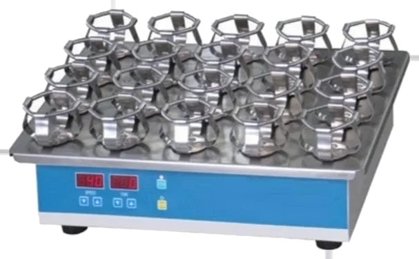
For mixing and development of cultures chemicals,solvents and assays etc in pathology,microbiology,chemical and bio technology.
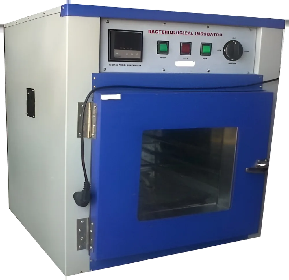
Best for: Incubators
Tell us what you are setting up and roughly how many of each. We will suggest a configuration and prepare one quote for the whole list.
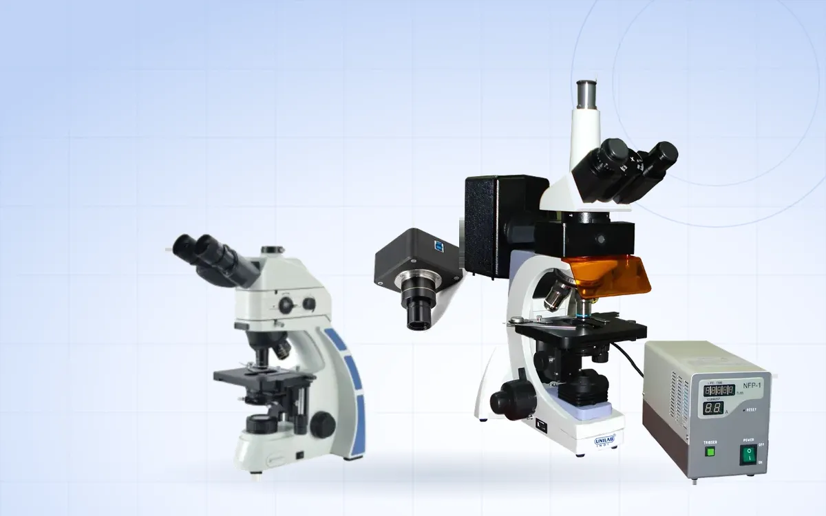
Filters, light sources, a dark room and a few working habits. A practical overview for laboratories adding fluorescence to routine microscopy.
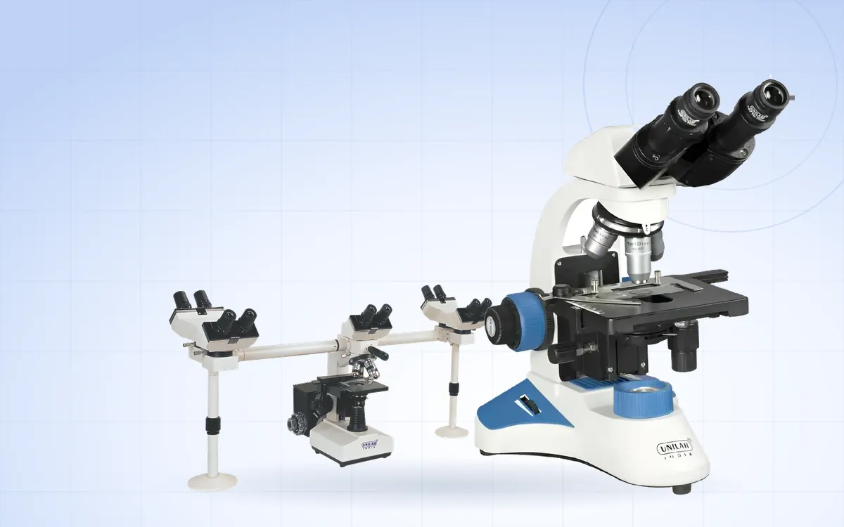
Even, bright and sharp. Setting up Köhler illumination takes about a minute and makes a visible difference to every slide you look at.
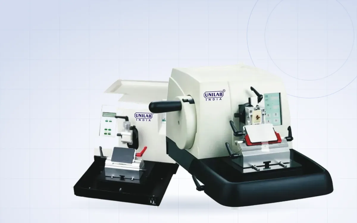
Thick and thin sections, chatter, crumbling ribbons. Most sectioning problems come down to block temperature, blade condition and a few machine settings.